The knee joint is made up of three compartments. Two of the compartments bear weight. The third compartment is the patellofemoral compartment, where the kneecap moves up and down when the knee bends and straightens. For the purpose of learning about MIPKR, let’s talk about the two weight bearing compartments, the medial, or inside compartment and the lateral, or outside compartment. These are shown in the diagram. The blue in the diagram represents cartilage. Cartilage in a knee joint is what coats the bone at the joint surface, it is several millimeters thick and when healthy it has a white shiny slick surface. With osteoarthritis the cartilage gets soft, develops cracks, pieces of it fall off, which act as an irritant to the knee. The end stage of this cartilage degeneration is when the entire thickness of that cartilage cushion is worn off exposing the underlying bone.
Bow Legged

Our knees are not all the same. Some of us, often more athletic types, have a bow-legged alignment and our weight mostly goes through the inside or medial compartment of the knee. Over time the cartilage may then wear out on that medial side, whereas on the lateral side the cartilage may be just like a teenager.
Knock-Knee Deformity
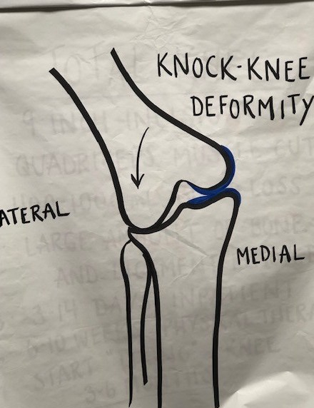
The opposite can also happen with those who are a little knock-kneed, the lateral compartment wears out. These alignment abnormalities causing one sided wear are by far the most common reason to develop osteoarthritis.
Wear of the patellofemoral joint can also occur by itself or in combination with wear of the weightbearing compartments. This usually causes more problems with getting up and down, squatting and kneeling. Many people can walk just fine on level ground even if the patellofemoral joint os worn bone against bone.
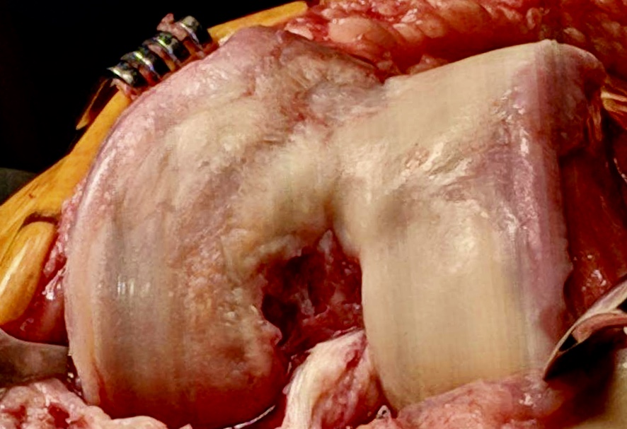
Here are some pictures of what unicompartmental arthritis looks like

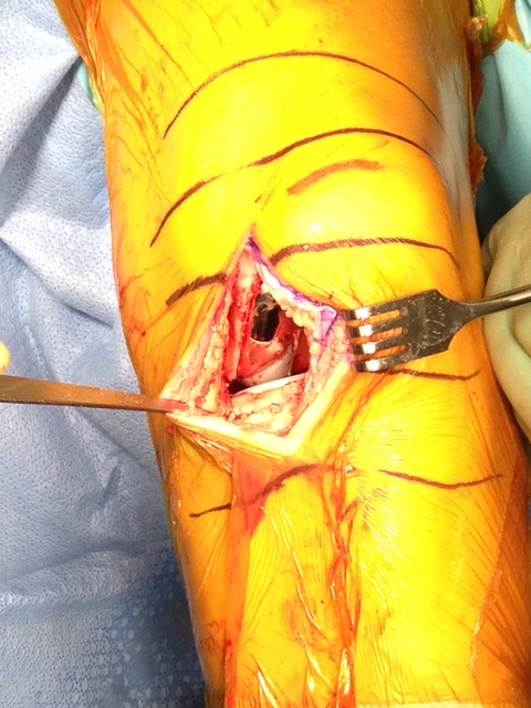
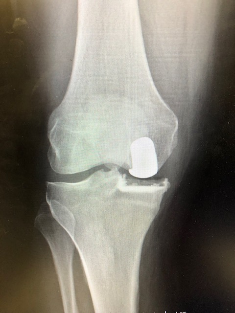
Here are some pictures of MIPKR